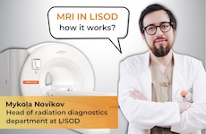
We asked Mykola Novikov the radiologist, head of the radiation diagnosis department LISOD, Ph.D. (Medicine) about MRI diagnosis and its features.
— Please, share with us what makes MRI technique special in diagnosis of malignant neoplasms?
Oncology uses a variety of imaging techniques to formulate an accurate diagnosis. These include ultrasound, mammography, CT, MRI, PET-CT, others. Choosing the technique depends on the clinical situation.
MRI allows us to see images of human’s body soft tissues, which sometimes is difficult to see using other techniques.
Having said that, I want to make it clear – the purpose of the MRI is not to detecting some very small lesion. It is about getting as much details as possible about what remains unclear after other imaging techniques were used.
Unlike the X-ray technique, MRI has no radiation exposure and does not use X-rays , it is rather based on a nuclear magnetic resonance.
— What cancer diseases can MRI detect?
This technique is most advantageous in neuroimaging, I mean diagnosing tumorous and non-tumorous illnesses of the brain. MRI is also indispensable in giving a picture of liver lesions, especially if hepatospecific contrast agents are used during imaging. In case of cancer patients, we are able to detect and interpret prostate lesions. MRI complements CT data in interpreting kidney lesions, it also works well for bone-marrow lesions in lymphoma.
MRI is also one of the diagnosis tools of breast diseases when for example, mammography or ultrasound imaging do not help much in understanding the nature of changes detected. Also, the technique is good to find out status of the breast implant. A biopsy can be MRI aided too.
On a separate note, in some countries MRI screening of the breast is advisable for the population with a high risk of breast cancer, Ukraine is not part of those first of all, due to socio-economic considerations (it is very pricy). Such approach however, exists in Israel for the Ashkenazi population, that has genetic factors causing early development of aggressive breast carcinoma in women. Also, currently in the USA an MRI breast screening for certain high-risk groups is being debated.
— Can MRI detect tumor metastases?
Sure, in certain scenarios we can see both the location of the primary tumor and tissues it spread to.
— What information about lesion MRI can provide?
MRI exam helps us understand the structure of neoplasm. Compared to computed tomography, MRI is more contrast, when we examine a tumor, not only we can see if it is dense or not, liquid or solid, with or without vessels, but much more than that, we see where the fat, the blood, or cystic components are. We can evaluate diffusion-weighted images, cell density. To sum it up, we get more details about lesion, and consequently, in some cases, can be more certain about formulating a diagnosis as opposed to the picture CT gives. Surely, in some case CT only is good enough.
However, when a CT does not give a clear-cut answer for example, in case of unclear or very small structures in the liver, magnetic resonance is very informative.
— What else except diagnosis MRI scan can do?
Because of the high contrast in soft tissues, one can be much more confident about the locoregional staging of a variety of tumors, whether intestine, or prostate, or certain oncogynecological diseases. Computed tomography does not have such a contrast between the tumor and tissue nearby, while magnetic resonance because of its physics, enhances this contrast. It becomes clearer where the tumor ends and some reactively changed tissues or healthy ones begin. This is very important for determining the T-values of staging, purposed to determine a method of treatment (e.g, surgery or radiation therapy).
MRI can be used for planning a radiation therapy. I’ll repeat myself, the technique is more precise in delineating boundaries of the tumor. It will work well in planning radiation therapy for head and neck tumors, pelvis, intestines, in neuro-oncology, in oncogynecology. MRI helps to determine tumor contour more precisely, the amount of radiation will be smaller (no irradiation exposure to healthy tissues), accordingly, the risk of negative side effects of radiation treatment will be less.
This is equally important in planning the surgery. Having an accurate imaging, we are more likely to understand whether adjacent structures are affected, those that are important for making a decision if tumors of the head and neck, cervix, bowel, liver, other are fit for surgery.
— What are the contraindications to using this technique?
Presence of ferromagnetic fragments in the body, which were implanted or accidentally got inside (sadly, many patients todate have injury caused fragments) is a strict contraindication for MRI exam. These are materials that can heat up and move in a high-intensity magnetic field. This is dangerous, first of all, for the patient during magnetic resonance imaging. This is a contraindication we have no control of.
There are also relative contraindications, such as claustrophobia. Some can get through the examination, and some can’t. In complex cases, sedation of the patient, is an option for example, in a state of psychomotor excitement. The MRI technique is highly sensitive to movement, therefore receiving a high quality image if the patient can’t lie still is difficult if not impossible.
Examination of patients with a very high body mass index is a challenge, as they may just not fit in tunnel of equipment, such cases are rare, though.
Examination during pregnancy is permitted, especially in the first trimester, method is used for medical reasons, and the second trimester is not a contraindication for diagnosing. I mean, the contraindication is relative, if making an exam is clinically important, anytime is good to make it.
— Does the exam require a special preparation?
It depends on examination. Some types of diagnosis don’t, some do. Patient will receive a guidance as they make an appointment.
— What MRI scanner LISOD has?
MAGNETOM Sempra 1.5 T magnetic resonance tomograph, a versatile device that suits well for every day clinical tasks, including in oncology. Undoubtfully, a 3 T devices can have certain advantages, but in most cases 1.5 T is quite enough. Furthermore, 1.5 T machine has better imaging capabilities versus more powerful equipment.
— How much time exam takes, what is the waiting time for results and how to make an appointment?
Time of diagnosis depends on area of examination. Sometimes it is 5-7 minutes, sometimes an hour. Same in case of result. Some patients receive it 15-20 minutes after, at times it may take 2-3 days to interpret.
Appointment is available via LISOD Contact Center at 0-800-500-110, calls in Ukraine toll-free.


 Dr. Meir Zahavi
 Dr. Sergey Baido
 Dr. Vadim Korpyak
 Dr. Аndriy Saulov