
Experience over 30 years
-
ABOUT HOSPITAL

The LISOD Hospital is a top grade multi-field hospital which is also a member of the European Organization for Research and Treatment of Cancer (EORTC, No. 2901) and associated member in other global cancer fighting organizations. The clinic has been active in Ukraine since 2007.
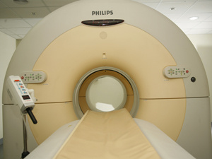
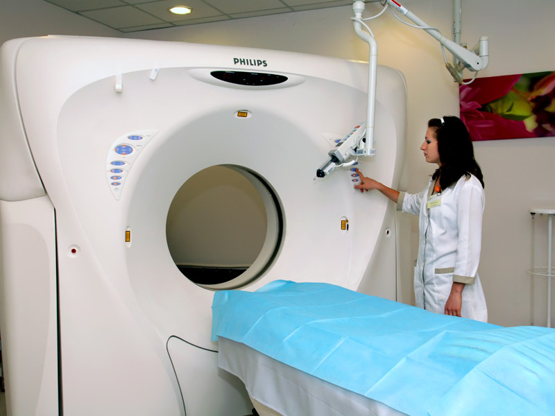
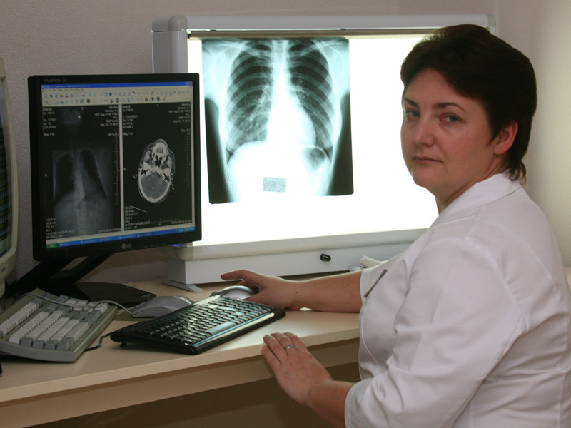
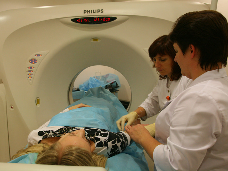
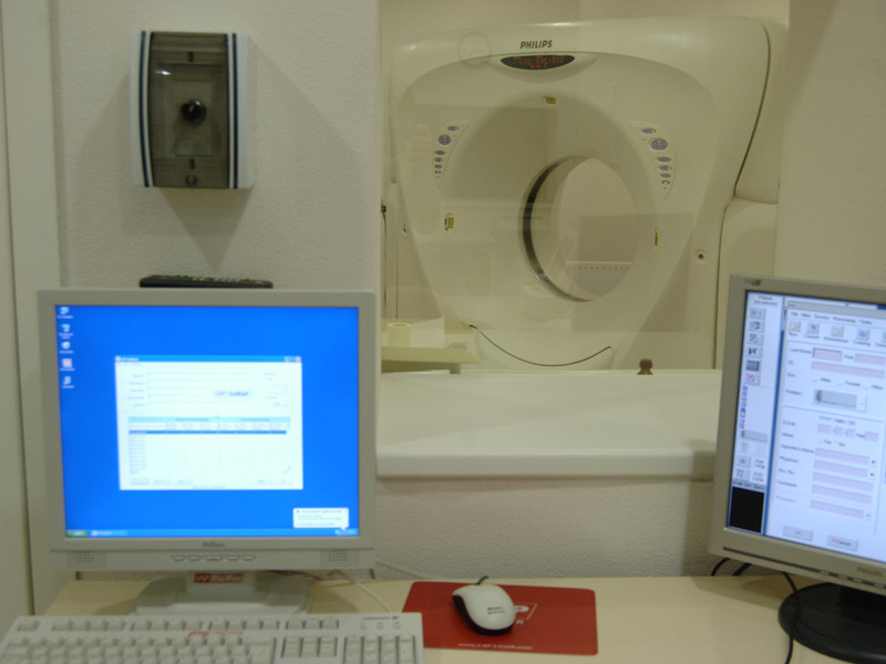
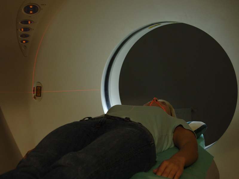
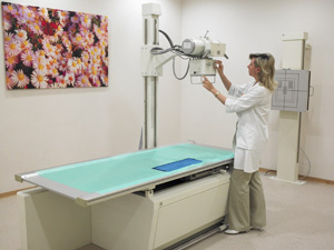
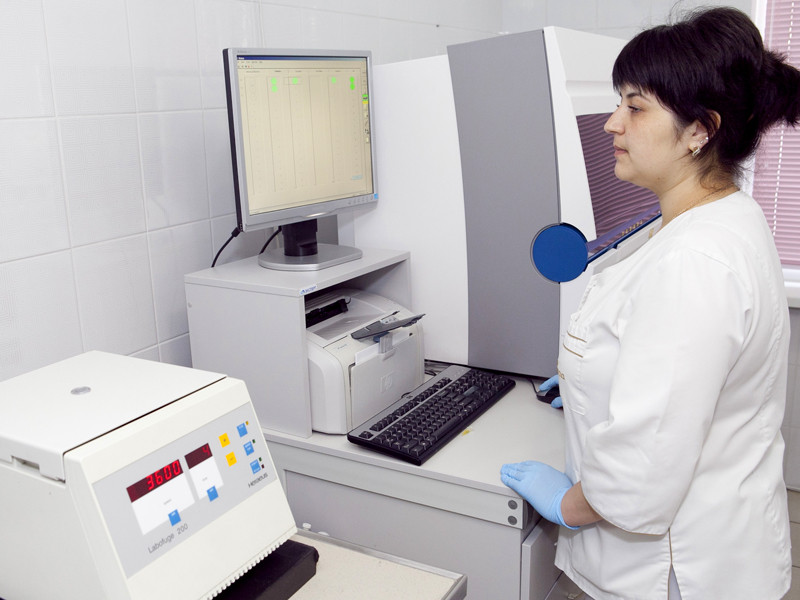
The Hospital has departments of radio diagnostics, clinical diagnostics, radiotherapy, chemotherapy, surgery and the Centre for Modern Mammology.
Close -
Our Experts

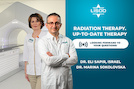
LISOD Chief Medical Officer
MD. PhD. Surgeon. Oncological gynaecologist. Doctor of Medical Sciences
 Dr. Meir Zahavi
 Dr. Sergey Baido
Leading surgeon in the CIS countries and Europe. Work experience spans over 20 years.
 Dr. Vadim Korpyak
Surgeon. Endoscopist.
 Dr. Аndriy Saulov
Clinical oncologist
Close -
Departments

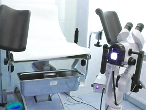
Since 2009, there has been a Centre of modern mammology at LISOD.

The vast majority of surgical interventions in LISOD Hospital are conducted using the laparoscopic method.
Close -
Cancer Treatment
- Mesothelioma
- Multiple Myeloma
- Myelodysplastic Syndrome
- Ovarian Cancer
- Pancreatic Cancer
- Acoustic Neuroma
- Rare Blood Disorders
- Skin Cancer
- Soft Tissue Sarcoma
- Bone Cancer
- Testicular Cancer
- Thymic Tumors
- Thyroid Cancer
- Breast Cancer
- Esophageal Cancer

We can comfortably and safely make the vaccination.

Comfortable and painless screening for one day.
Close -
Education and Training

Chief activities of the LISOD Hospital include not only cancer treatment and fight in general, but also introduction of up-to-date technologies and methods of treatment. We are always open for cooperation with our colleagues from Ukraine and abroad.
Close